False Positives/Negatives
CT scanning is highly sensitive for thymomas. If the thymus appears grossly asymmetrical or if it has a lobular configuration, the diagnosis of a thymoma should be strongly considered. However, thymomas cannot be distinguished from other thymic masses on CT scans unless fat is visible. Thymomas are usually homogeneous and show mild enhancement with the use of contrast. They tend to grow to one side of the mediastinum or the other. (Images of thymomas appear below.) [2]
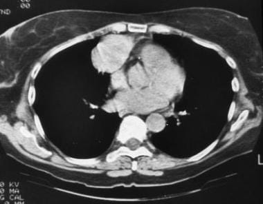
 Malignant thymoma. Chest CT scan in a 61-year-old man with myasthenia gravis demonstrates a large, lobulated mass with punctate calcification in the anterior mediastinum; these findings are consistent with a thymoma.
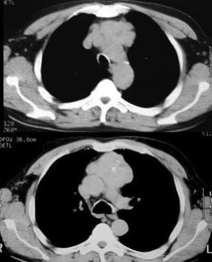
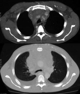 Invasive thymoma. Contrast-enhanced CT scan shows a thymic mass with variable attenuations invading the adjacent mediastinum. Bilateral pleural effusions are also present, indicating pleural invasion.
When cystic degeneration, necrosis, and old hemorrhage are present, the affected areas are of low attenuation. Normal thymus that has undergone fatty infiltration and a pulmonary mass adjacent to the anterior mediastinum sometimes mimic a thymoma. In cases of encapsulated thymoma, CT scanning often reveals a preserved fat plane completely surrounding the mass. Fibrous adhesions and inflammation may mimic invasion of the tumor.
On CT scans, invasive thymoma is often characterized by encasement of mediastinal structures and pericardial/pleural implants; in advanced cases, transdiaphragmatic spread is seen.
Comments
Post a Comment