Thymic Lymphoma
On CT scans, the thymus is enlarged, symmetrically or asymmetrically; this enlargement often makes it difficult to differentiate the thymus from the enlarged surrounding lymph nodes. At times, differentiating the thymus from a thymoma is difficult, although the clinical picture and the presence of other sites of lymphadenopathy are often helpful in the diagnosis. (See the image below.)
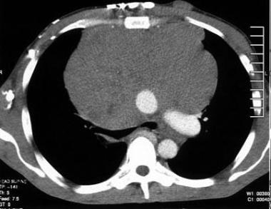 Thymolymphoma. Contrast-enhanced CT scan demonstrates a large, lobulated, soft tissue mass in the anterior mediastinum that posteriorly displaces the aorta and pulmonary artery.

Comments
Post a Comment