Thymic Lesion
Overview
Thymic tumors occupy the anterior mediastinum, which is immediately posterior to the sternum and the anterior surface of the pericardium and great vessels. Tumors of thymic, lymphatic, or germ cell origin most commonly occur in this compartment, although aberrant parathyroid or thyroid tissue masses are sometimes found, along with vascular and mesenchymal tissue masses. (See the images below.)
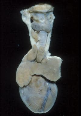
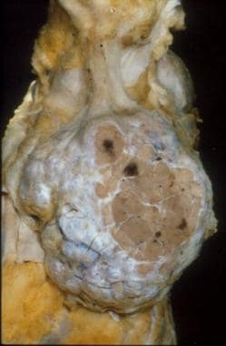 Gross appearance of a thymoma showing distinct multinodularity and scattered, focal cystic changes.
About one half of all thymic tumors are malignant in individuals aged 20-40 years, and one third are malignant in persons younger than 20 years and those older than 40 years. (A malignant thymic tumor is seen in the image below.)
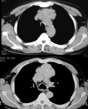 Malignant thymoma. Chest CT scan in a 61-year-old man with myasthenia gravis demonstrates a large, lobulated mass with punctate calcification in the anterior mediastinum; these findings are consistent with a thymoma.
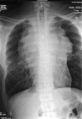
Approximately two thirds of all mediastinal tumors and cysts are symptomatic in children, whereas only a third are symptomatic in adults. When all age groups are considered, nearly 55% of patients with benign mediastinal masses are asymptomatic at presentation, compared with only approximately 15% of those in whom masses are found to be malignant. Computed tomography (CT) scanning is most valuable in the diagnosis of thymic lesions and anterior mediastinal masses. Magnetic resonance imaging (MRI) may also be used. [1, 2, 3, 4, 5, 6, 7, 8]
Comments
Post a Comment