Radiography
Many thymomas may be visualized using routine chest radiographs in posteroanterior (PA) and lateral views. A thymoma frequently appears as a smooth, lobulated mass in the superior aspect of the anterior mediastinum, often projecting into one of the hemothoraces. The mass may be calcified or cystic. (See the images below.)
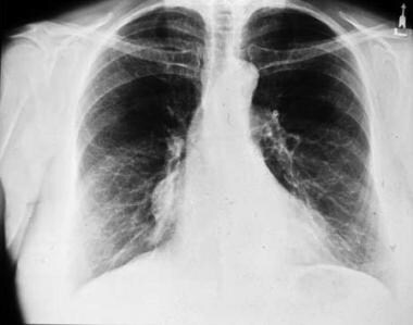 Low-lying thymoma. Plain chest radiograph shows a mass lying against the right heart border.
In most cases, thymolipomas are detected incidentally on chest radiographs. They are often large, ranging up to 36 cm in diameter (mean, 18 cm) at the time of diagnosis. Thymolipomas may project into the hemithoraces; they may be seen draping over the heart and extending as far as the costophrenic angles. They can be easily mistaken for pleural or pericardial tumors or even pulmonary sequestration.
Comments
Post a Comment