Thymolipoma
CT scanning reveals a fatty mass interspersed with varying amounts of thymic soft tissue. The whole mass often consists of adipose tissue except for a thin rim of thymic tissue; this finding is consistent with soft tissue attenuation and mimics a pure lipoma of the mediastinum. (See the image below.)
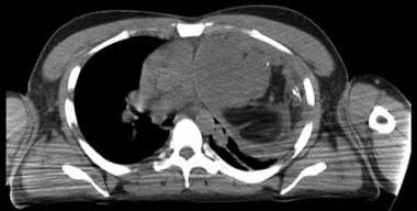 Thymolipoma. CT scan of the chest shows a large anterior mediastinal mass displacing the mediastinal vascular structures to the right and projecting into the left thorax. The tumor is composed of soft tissue and fat, and a few punctate calcifications are present.
Comments
Post a Comment